Treatments
Guide
Diagnose Yourself
Disclaimer: Patient education is very important, however this site is a guide to help patients understand their condition and does not replace years of training and clinical experience.
Make a Diagnosis
Before an appropriate treatment can be instigated, first and most importantly, one must make the correct diagnosis.
Often this can be achieved by simply taking a history and doing an examination.
To aid in the diagnosis, we can use radiology (x-rays, CT, MRI, ultrasound, etc). Each of these modalities is used to look for different things. Depending on the condition, it would be incorrect to use some of these modalities for a given condition, as it may not help the diagnosis and may place patients in harm's way, for example radiation from an x-ray or CT scan for a soft tissue concern.
A doctor will usually start by making a differential diagnosis, a list of possible diagnoses ordered from most likely to least likely. This, however, is only possible if you already know the whole list of diagnoses. For a medical student, or someone without a medical background, a different approach is needed. Anatomically is the best method.
Identifying the pathological anatomy is the most important factor when using this easy technique. At first, you don't need to know what exactly the anatomy is, you just need to identify where the anatomy is. One must pay very close attention to exactly where the problem is, or the biggest problem, e.g. the most pain. Normally there will be one area of pathology causing many, or diverse, symptoms, as opposed to many discrete areas of pathology.
Step 1: Identify the Problem
- Is it pain?
- Clicking?
- Catching / locking?
- Stiffness?
- Numbness / tingling?
- Weakness?
- Swelling / deformity?
- Other?
Pain: Sometimes it's hard for people to exactly localise their pain, it may feel that it's around a general area, or radiates to different places. This is very common. It's best to try and find the site where the pain is the worst, ideally being able to put one finger on the worst spot. The more spots of "the worst" pain, the harder it is to make a diagnosis and give the best treatment. If you look hard enough, you will often find one spot where it is the worst.
Clicking: Some joints click and are not painful, so usually don't need any treatment and won't cause any long-term problems. Painful clicking, however, needs further evaluation. If one can identify the exact point of clicking, or where the pain is when it clicks, this is ideal.
Catching / Locking: This is usually a very specific symptom, and depending where it is in the body, it can be very easy to make a diagnosis.
Stiffness: This is a very common symptom, especially after an injury. It's also one of the first signs of arthritis, although people may not know that their joint has a decreased range of motion. Luckily, most joints have an opposite joint with which you can compare. If your hand is stiff, try to identify exactly which joint(s) are stiff.
Numbness / Tingling: This will normally occur in the distribution of a sensory nerve. Identifying exactly where the numbness is, is vital, exactly which fingers. Commonly people say "it's my whole hand", normally because they have not paid acute attention when the symptoms are present. During the symptoms, pay distinct attention to exactly which fingers, or sides of fingers, are involved.
Weakness: This can be a vague symptom and has many possible diagnoses. It's important to figure out exactly what is weak. Find specific tasks that you find difficult; is it pain that causes the weakness, or does it just not work?
Swelling / Deformity: These are common all over the body and can usually be pointed out to your doctor very easily and diagnosed quickly. Swellings in certain areas are characteristic of certain conditions.
Other: Occasionally one cannot put a label on what is wrong, it just doesn't feel right, it feels unstable, it gives way, etc. Again, you can still identify more information if you really concentrate on anatomy and exactly when it is a problem. "My wrist gives out when I go to the gym" is less helpful than "my wrist gives out secondary to pain, when I am lifting a dumbbell at the gym, with my elbows at right angles with palms facing together, and I deviate my wrist to the little finger side, ulnar deviate".
Step 2: Identify What the Relevant Anatomy Is in the Area
Learning the entire anatomy of the body takes years. If you are focusing on a specific area of a problem, then you can often learn the required anatomy in minutes. Below are common sites of pain.
Hand and Wrist
"A disease has not read the description of itself"
– a condition does not always present in the same way in every patient.
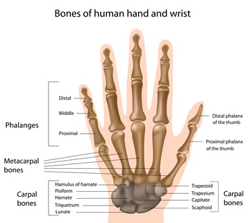
The picture below shows the bone anatomy of the hand. Any bone can be fractured, or the joints between can become arthritic.
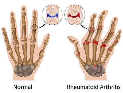
Arthritic joints often start by having swelling and a decreased range of motion, plus or minus pain. This can progress to deformity and/or dislocation of joints.
By identifying certain areas that have characteristic spots that cause pain, these painful areas are usually quite discrete. Arthritic processes such as a SNAC or SLAC wrist, mid-carpal instability, or rheumatoid arthritis can present with global pain around the wrist.
Dorsal Wrist Pain
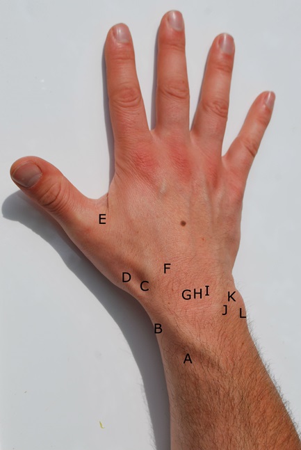
A = Intersection syndrome
B = De Quervain's tenosynovitis
C = Scaphoid fracture / Wartenberg's syndrome (nerve entrapment)
D = Base of thumb arthritis
E = Ulnar collateral ligament injury
F = Carpal boss
G = Scapholunate ligament tear
H = Kienböck's disease
I = Lunotriquetral ligament tear
J = DRUJ (distal radioulnar joint) arthritis / instability
K = Ulnar impaction / TFCC tear
L = ECU dislocation / tendonitis
Volar Wrist
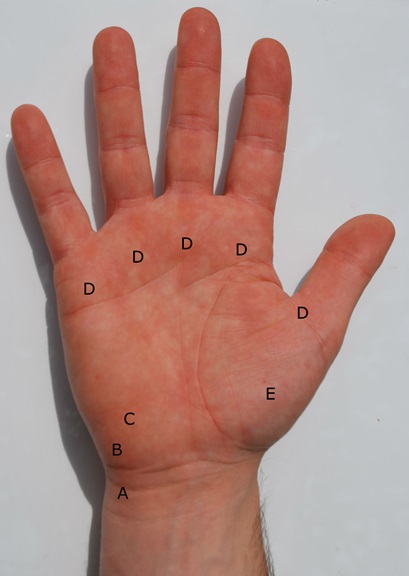
A = FCU tendonitis
B = Pisotriquetral arthritis / ulnar nerve entrapment / ulnar artery thrombosis
C = Hook of hamate fracture
D = Trigger finger
E = Base of thumb arthritis
Condition
Hands
Hand and wrist conditions cover a wide range, from acute soft tissue injuries to nerve compression and the therapy that supports recovery from both. Select a topic below for more detail.
Arthritis is very common, wear and tear upon a joint that presents as pain and stiffness. The most common causes of a joint wearing out are trauma and genetics. Some people will fracture into a joint, and that joint is then likely to develop post-traumatic arthritis at some point. With genetics, though, some people will never develop arthritis well into their 90s, while others' joints wear out in their 30s, or even younger.
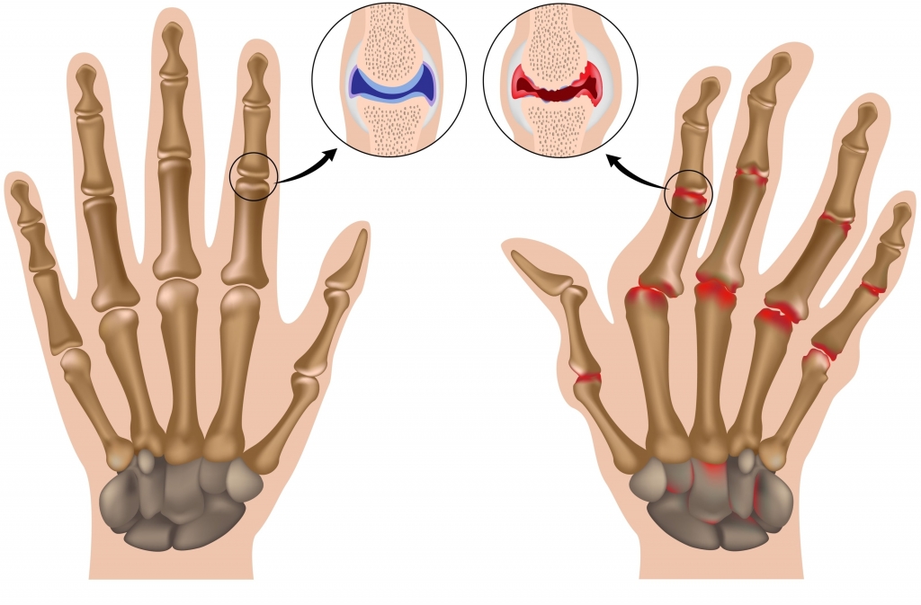
Osteoarthritis
Very common, and can affect any joint, with the small joints of the hand being particularly prevalent. Therapy and anti-inflammatories are the best initial treatments, with splinting appropriate for certain joints, namely the base of the thumb. If and when these measures fail, function and pain relief become the primary goals. Depending on the joint, excision, replacement, or fusion may be the best option, tailored to the individual's needs, as some treatments maintain range of motion at the expense of power, while others maintain power at the expense of range of motion.
Base of Thumb Arthritis
One of the most common arthritic conditions in the body, occurring across most age groups but most commonly affecting middle-aged women. It starts with pain at the base of the thumb, on either the palm or back side, along with a loss of the thumb's normal contour. Patients often notice problems opening jars, turning door handles, or with strong gripping motions.
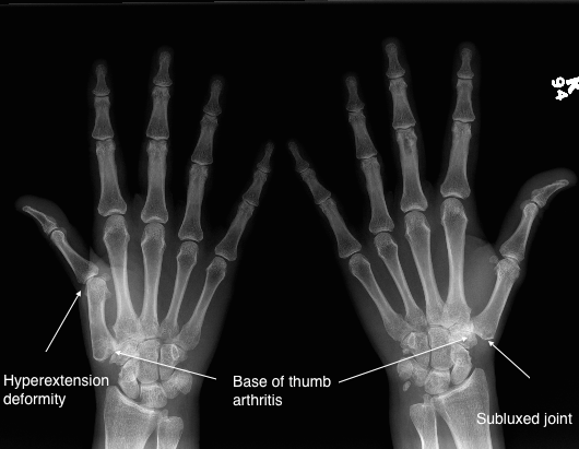
For more information: Base of Thumb Arthritis.
Finger Arthritis
Any of the joints in the fingers can become arthritic, often across multiple digits. Treatment depends on exactly which joint is involved and what the individual patient wants: replacement is a good option for some joints, while fusion suits others better.
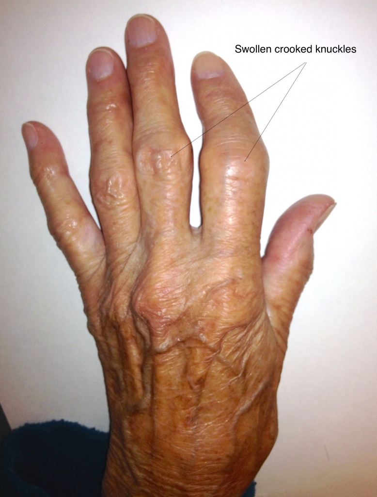
For more information: Arthritis of the Hand.
Scapholunate Advanced Collapse (SLAC)
This pattern of arthritis is the natural history of a torn scapholunate ligament in the wrist. The initial injury is often fairly benign, but the wrist is never quite right afterwards, and the process typically takes a decade or more to develop. It occurs because abnormal wrist kinematics let the bones move independently, causing abnormal stresses that lead to arthritic degeneration; occasionally it can be symptom-free until the arthritis develops. Treating the underlying injury acutely is the best way to prevent this process starting, using either an anatomic or non-anatomic surgical reconstruction. As the arthritis progresses, an excision, a Proximal Row Carpectomy (PRC), or a partial (four-corner) fusion may become the more appropriate option.
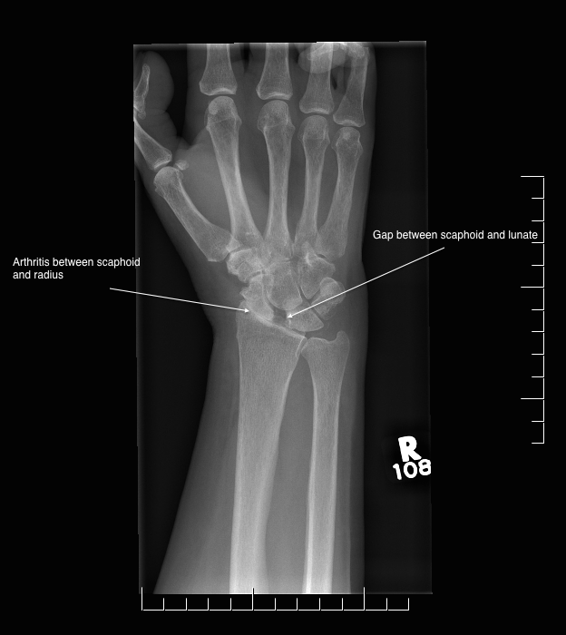
SLAC wrist follows a predictable pattern of progression, from the radial styloid, to the whole radio-scaphoid joint, and lastly to capitolunate arthritis.
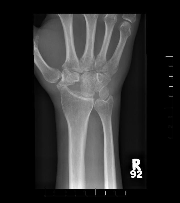
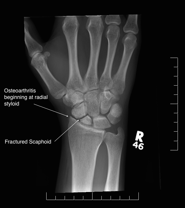
Scaphoid Non-Union Advanced Collapse (SNAC)
This pattern of arthritis is the natural progression of a scaphoid fracture left untreated, very similar to a SLAC wrist in its cause, presentation, and treatment. As with a SLAC wrist, the lunate is no longer held in a fixed position, so it starts to tilt backwards (dorsally), leading to a DISI (dorsal intercalated segment instability) deformity that reflects the altered kinematics of the wrist.
Carpal Boss
A carpal boss is an enlargement of the bone at the base of the second or third metacarpal. It's a firm swelling, often mistaken for a ganglion. Treatment is splinting and/or steroid injections and anti-inflammatories; operative debridement is rarely needed.
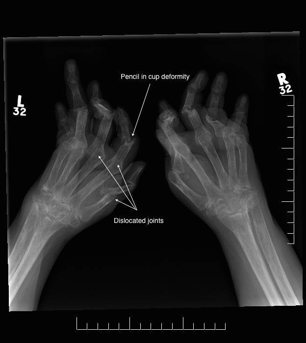
Rheumatoid Arthritis
The mainstay of treating rheumatoid arthritis is non-surgical, supervised by a rheumatologist. There does come a point, though, when function starts to deteriorate due to tendon rupture, deformity, or loss of function. Surgery is often very successful in rheumatoid patients and can restore significant lost function; it's important not to wait too long before having surgery for this condition, as leaving it too late can shift the goals of surgery from regaining function to simply stopping progression, due to soft tissue and bony destruction.
Surgery can achieve many different things, from making the hand look and align like a hand again, to something as small as relocating dislocated tendons, through to repairing ruptured tendons or a total wrist fusion or replacement. Since rheumatoid arthritis often involves many joints, the exact combination of treatments differs for each person.
Nerves are very sensitive to compression. When a nerve is compressed at any point along its length, it can produce symptoms further down, numbness, tingling, pins and needles, or a "dead" feeling. Simply relieving the pressure often resolves the problem, though the degree of recovery depends on several factors, particularly how long the symptoms have been present.
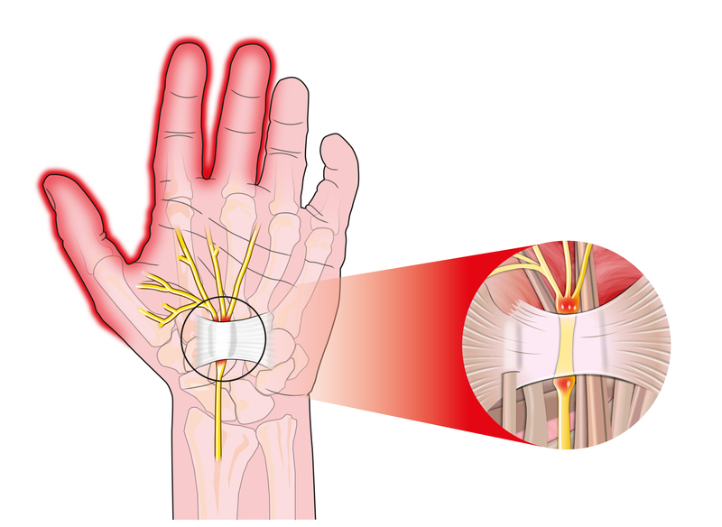
Median Nerve (Carpal Tunnel Syndrome)
Usually presents as numbness and tingling in the thumb, index, middle, and half of the ring finger, often starting at night or during specific activities. Left untreated, this can progress to constant numbness, muscle wasting, and weakness. Initial treatment is nocturnal splinting; if this doesn't resolve it, a straightforward surgical release of the ligament across the wrist is very effective.
Ulnar Nerve (Cubital Tunnel Syndrome / Guyon's Canal)
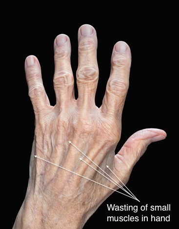
Less common than carpal tunnel syndrome, presenting as numbness and tingling in the little finger and half of the ring finger, sometimes affecting the palm depending on where the compression occurs. It can affect the small muscles that spread and cross the fingers, and in chronic cases these muscles can visibly waste away. Compression most commonly occurs at the elbow, less often at the wrist. Surgical release is very successful where non-surgical management hasn't worked.
Radial Nerve
The radial nerve doesn't supply any muscles in the hand, only sensation. Compression can cause tingling on the back of the hand between the thumb and index finger, or a dull ache at the back of the wrist from compression near the elbow. Therapy is usually effective, and surgery is not commonly required.
Osteonecrosis is an unusual condition where the blood supply to a bone stops and the bone dies. There are a number of modifiable risk factors that treatment aims to address.
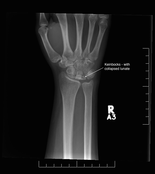
Kienböck's Disease (Avascular Necrosis of the Lunate)
Associated with having a relatively short ulna. Treatment depends on the degree of symptoms present, and even quite advanced arthritis can sometimes cause relatively few symptoms.
For more information: Kienböck's Disease.
Preiser's Disease (Avascular Necrosis of the Scaphoid)
Much less common than Kienböck's disease. Non-operative management with splinting and oral medication is often successful; where this doesn't resolve it, a SNAC wrist procedure gives good results.
Soft tissue injuries of the hand and wrist cover a range of ligament and cartilage conditions that can be easily missed on initial assessment, particularly when x-rays appear normal. Below are some of the more common conditions seen and treated.
Scapholunate Tear
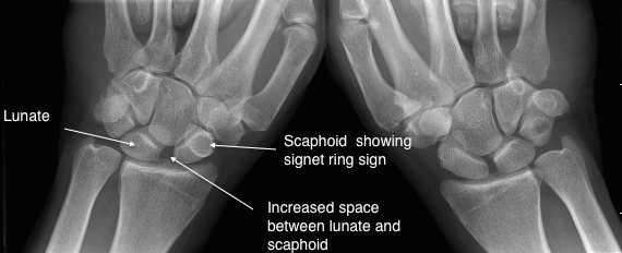
Often misdiagnosed as a simple wrist sprain. Standard x-rays can look normal unless stress views are taken (imaging the wrist while making a fist), which can reveal widening between the lunate and scaphoid, along with a rounded outline in the scaphoid indicating it is flexing separately from the lunate. Left untreated, this injury will typically progress to a SLAC wrist over roughly a decade. Common symptoms are a dull ache in the wrist and a sensation of the wrist giving way. Acute tears are generally treated surgically.
Lunotriquetral Tear
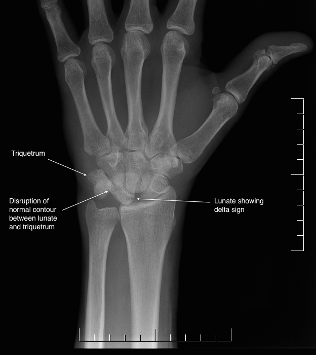
Less common than a scapholunate tear, but carries the same long-term risk of arthritis from disrupted wrist mechanics. Presentation is similar, pain around the wrist, weakness, and a sense of the wrist giving way. X-rays may show the "delta sign" (a triangular-appearing lunate) and loss of the normal Gilula's arcs, with lateral views showing a VISI deformity (Volar Intercalated Segment Instability). Treatment is surgical stabilisation.
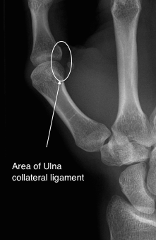
Ulna Collateral Ligament Injury (Thumb)
Commonly known as skier's thumb when it occurs acutely, or gamekeeper's thumb when it develops chronically. The ulna collateral ligament stabilises the thumb during pinch and grip. A tear causes pain and instability when the joint is loaded. Initial treatment is bracing to allow healing; in cases that don't settle, surgical reconstruction of the ligament may be required.
TFCC Injury and DRUJ Instability
The Triangular Fibro-Cartilage Complex (TFCC) stabilises the distal radius and ulna, playing a role similar to the meniscus in the knee. Certain anatomical variations can increase the risk of injury, for example, when the ulna is longer than the radius ("ulna positive"), increasing impingement against the wrist bones. Treatment typically starts non-operatively, with splinting and anti-inflammatories. An MRI helps visualise the TFCC, as it isn't visible on plain x-ray, and arthroscopy remains the gold standard for diagnosis. Where the ulna is positive, an ulna shortening osteotomy, tailored to the individual patient, may be recommended. TFCC tears unrelated to ulna length can also be addressed arthroscopically.
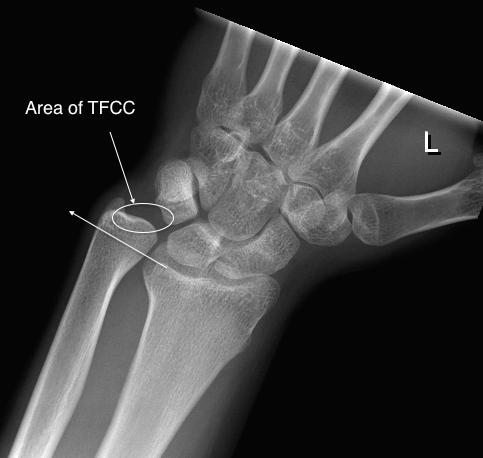
Area of the TFCC.
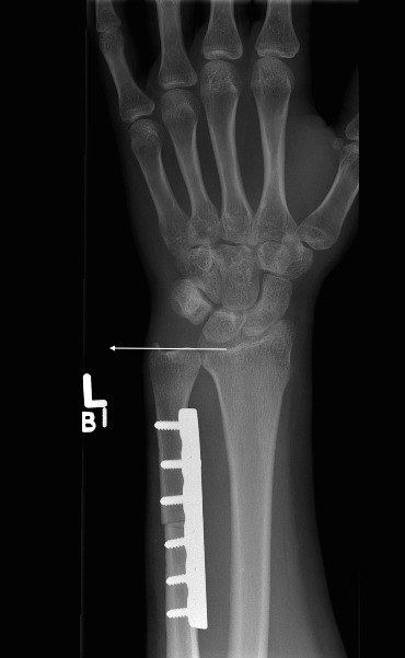
DRUJ instability.
Soft tissue pathologies of the hand, wrist, and elbow cover a wide range of common conditions affecting tendons, fascia, and joint linings. Below are some of the more common conditions seen and treated.
Dupuytren's Disease
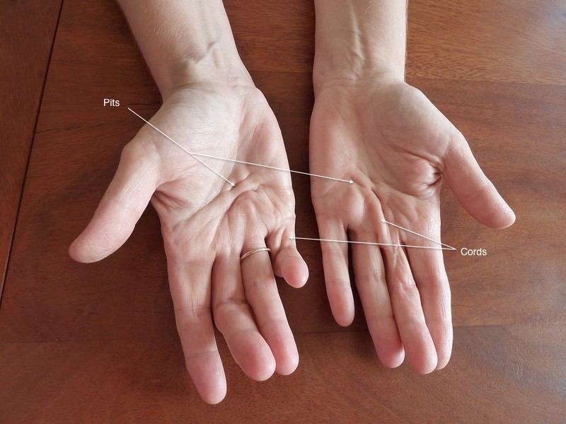
A very common genetic condition, thought to originate from the Vikings of old northern Europe. It's a disease of the tissue just under the skin, causing pits and lumps in the palms and fingers, and in advanced stages, curling of the fingers downwards. Splinting and other non-operative management won't cure or slow the condition, and it's important not to intervene too early, as operating before it becomes a problem can make things worse. The right time to treat is once it becomes a functional problem, for example when it starts poking you in the eye while washing your face, or you can no longer put your hand in your pocket. Treatment options range from needle fasciotomy and enzyme injection through to complete surgical excision, chosen depending on severity and the patient's wishes. Recurrence is higher with the less invasive treatments, though recovery is much quicker.
There are two related conditions involving the same thickening of connective tissue elsewhere in the body: Ledderhose disease, affecting the soles of the feet, and Peyronie's disease, affecting the penis.
For more information: Dupuytren's Disease.
Trigger Finger
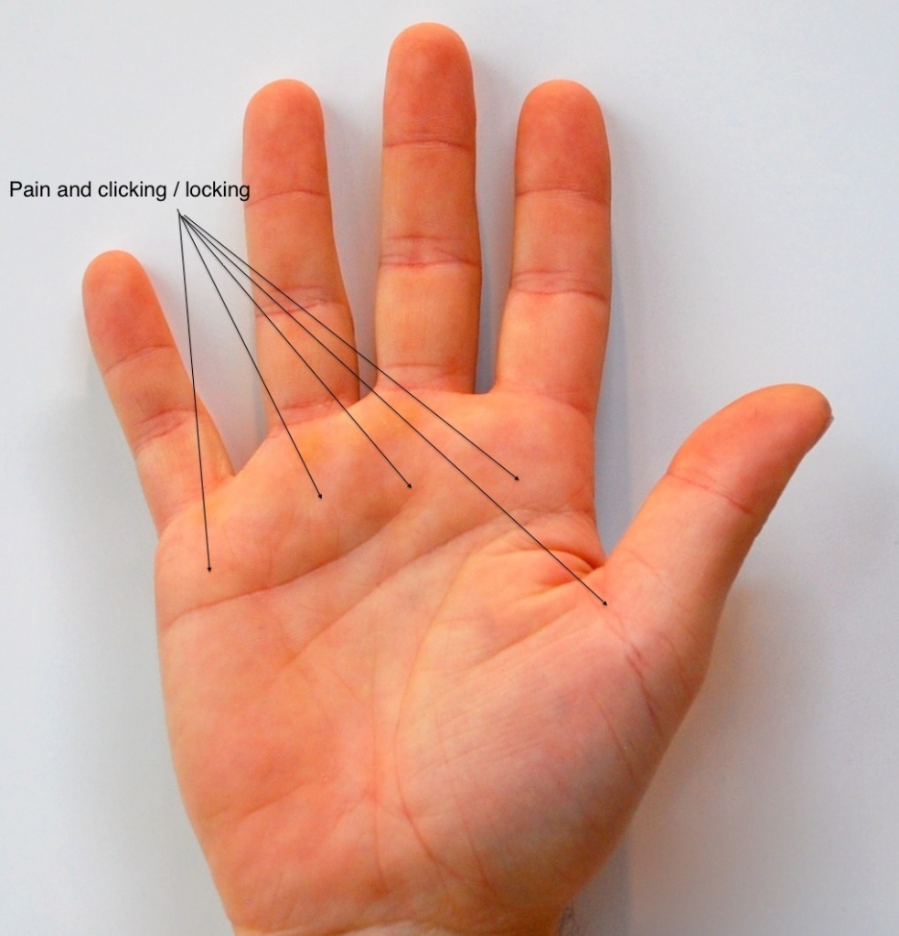
A very common condition presenting with pain in the palm near the base of the fingers, at the A1 pulley over the MCP joint. Thickening of the A1 pulley narrows the entrance to the flexor sheath, and the flexor tendon can catch as it passes through, causing the finger to click, or lock, as it's extended, sometimes needing help from the other hand to straighten. Occasionally there's a palpable nodule in the tendon, though this is much less common than textbooks suggest. Treatment starts non-operatively, with a steroid injection and nocturnal splint, curing 60-70% of cases. A second injection, if needed, has a similar success rate. If this also fails, surgical release is performed. Patients with multiple trigger fingers, or with diabetes, are less likely to respond to non-surgical treatment.
De Quervain's Tenosynovitis
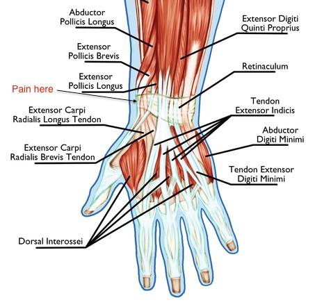
Inflammation of the first dorsal compartment tendons at the wrist, extensor pollicis brevis (EPB) and abductor pollicis longus (APL), which run along the thumb side of the wrist. It causes pain with thumb and wrist movement, particularly gripping or twisting motions. Treatment is typically splinting, anti-inflammatories, and a steroid injection; surgical release of the compartment is reserved for cases that don't settle.
For more information: De Quervain's Tendinosis.
Intersection Syndrome
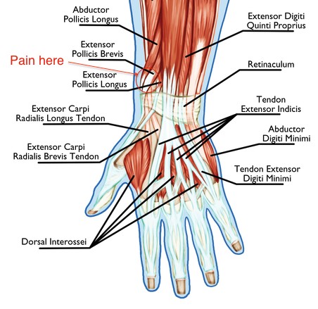
Another tenosynovitis, similar to De Quervain's but much less common, caused by friction where the first dorsal compartment tendons cross the second compartment tendons (ECRL and ECRB) as they travel through the forearm. It's most commonly caused by repetitive wrist movement, particularly making a fist and bending the wrist, and typically presents with pain and swelling a few centimetres up from the wrist on the back of the forearm. It rarely needs surgery, and usually responds to a splint and modification of activities.
ECU Instability
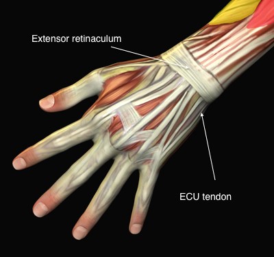
The extensor carpi ulnaris (ECU) is a muscle that extends the wrist, moving it backwards. Its tendon, along with the other tendons that extend the fingers, thumb, and wrist, passes under the extensor retinaculum, a fibrous band that holds the tendons in place. The ECU tendon can become loose and dislocate around the head of the ulna, which can become symptomatic. Anti-inflammatories and a brace can often minimise symptoms; if this fails, the tendon can be stabilised surgically using a small slip of the extensor retinaculum.
Mid-Carpal Instability
More common than previously recognised, presenting as vague wrist pain and weakness, often in young women who are ligamentously lax (double-jointed). It's a dynamic condition that can only be appreciated on physical examination; in most people, the mid-carpal joint can be subluxed (partially dislocated) without pain, but in this condition the same movement recreates the presenting pain. Treatment starts with non-operative management: splinting, steroids, and activity modification. This condition is self-limiting and tends to improve with time, though a minority of patients benefit from surgical tightening (plication) of the wrist capsule.
Tendonitis
There are many tendons around the wrist, any of which can become tender and inflamed. Patients can often identify this themselves, as the pain sits along a palpable tendon. Splinting, steroids, anti-inflammatories, and activity modification will usually help, depending on the specific tendon involved, and surgery is rarely required.
Tennis / Golfer's Elbow
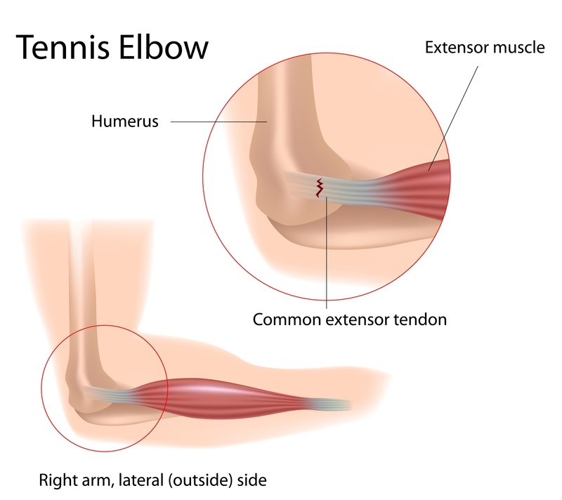
Pain on the outside of the elbow (tennis elbow) or inside of the elbow (golfer's elbow) are common conditions, causing localised pain around the bony prominences of the elbow. Despite the name, this is a degenerative change within the tendon just off the bone rather than true inflammation. Steroids, anti-inflammatories, and bracing will treat most cases, though surgical debridement can be performed for those that don't settle.
For more information: Tennis Elbow.
Ganglion
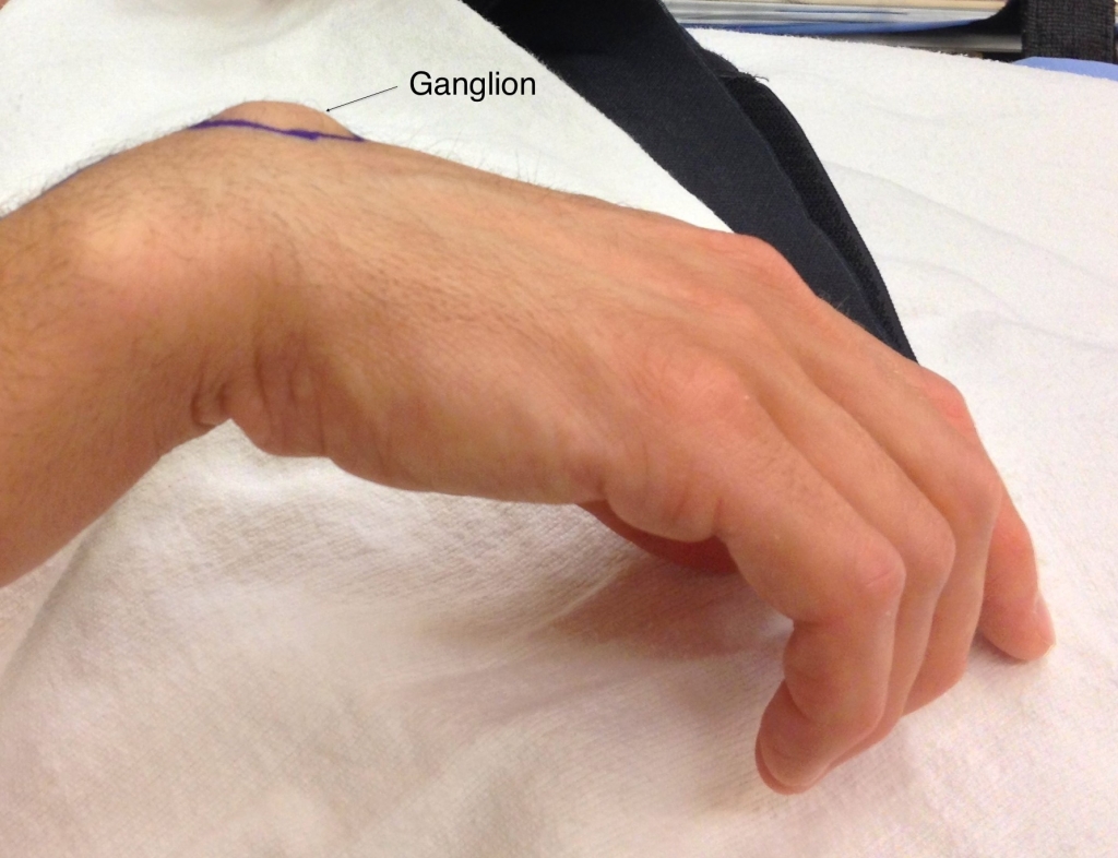
The most common "tumour," or swelling, seen in the hand. Ganglions are not cancerous and not dangerous, they're a fluid-filled outpouching of a joint or tendon sheath. Their size can fluctuate, and they can disappear on their own from time to time. They commonly occur around the back of the wrist, the underside of the wrist, or the tips of the fingers, where they're known as mucous cysts, and can also occur along the tendons of the fingers. Classical treatment was hitting it with a large book (the bible), though this had a high recurrence rate. Today, if it isn't bothering you, the best option is to leave it alone. If it's painful, restricts activity, or is unsightly, it can be aspirated (drawn out with a needle) or removed surgically. Aspiration has a high recurrence rate; removal also carries some risk of recurrence, though this is much less likely.
For more information: Ganglion Cyst.
Therapy is a core part of treating hand conditions, whether or not surgery is involved. Physiotherapists and occupational therapists support the non-operative, pre-, and post-operative management of most hand conditions. Any injury, surgery, or period of immobilisation, especially in the hand, can lead to stiffness, which can significantly affect a person's ability to function normally. For this reason, care is delivered as a team, involving the patient, surgeon, and therapists together.
Complex Regional Pain Syndrome (CRPS)
A poorly understood condition that affects the hand more commonly than other parts of the body, presenting as pain out of proportion to the original injury, sometimes following surgery or even a minor knock. The skin can take on a dull, mottled appearance, and normally non-painful contact, such as light stroking, can cause significant pain. Treatment centres on early, consistent therapy alongside medication, the sooner this starts, the better. Most cases resolve within about 18 months, though beyond 2 years the nervous system's response can become more difficult to reverse, which is why early, active treatment matters. Deep tissue massage, although intensely painful in the short term, doesn't cause any tissue damage, just as it wouldn't in someone without CRPS, and is one of the best ways to recover; patients can carry this out themselves. Protecting or avoiding use of the affected area tends to make outcomes worse, not better, and is much more likely to lead to long-term problems.
For more information: Complex Regional Pain Syndrome.
Wrist Arthroscopy
A surgical procedure used to both diagnose and treat wrist conditions, particularly useful when the diagnosis isn't clear from imaging alone. A small (2.7mm) fibre-optic scope is inserted through tiny incisions at the back of the wrist, allowing direct visualisation of the bones and soft tissues, often with treatment carried out at the same time, or the pathology identified for further discussion in clinic.
For more information: Wrist Arthroscopy.
Wrist Arthroplasty
An option for significant wrist arthritis where other treatments haven't helped, offering an alternative to wrist fusion. It's far less common than hip or knee replacement, and correspondingly less well studied, so its long-term longevity isn't as well established and at this point may be considered experimental. The main benefit is preserved range of motion; the trade-off is uncertainty about how long it will last. If it does fail, conversion to a fusion remains possible, though outcomes aren't quite as good as a fusion performed as the first procedure.
For more information: Wrist Arthroplasty.
Carpal Bones
Hook of Hamate Fracture
An uncommon bone to fracture, usually from repetitive impact rather than a single traumatic event, common in sports involving a bat or stick, such as baseball or hockey, and in New Zealand, cricket. It presents as pain at the base of the palm in line with the little finger, difficult to see on standard x-rays, a carpal tunnel view or CT scan is often needed. Treatment starts with stopping the causative activity; if that isn't possible or doesn't resolve it, the hook can be excised through a small incision in the palm.
Scaphoid Fracture
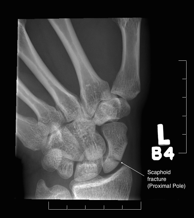
A very common fracture, typically resulting from a fall onto an outstretched hand. The scaphoid is particularly vulnerable since it spans from the proximal row of carpal bones across much of the distal row. Most common in young adults, it's uncommon in older people, whose weaker bones make a distal radius fracture more likely instead.
Bite Wounds
Fight bites are both common and can be particularly problematic. A simple tooth wound over the knuckle can become infected very quickly, and without early, aggressive treatment, can lead to multiple operations and significant morbidity.
For more information: Scaphoid Fracture, Finger Fractures, Hand Fractures, Distal Radius Fracture, Thumb Fractures, Human Bites.
Treatment
Hip Replacement
Arthritis is the gradual wearing out of a joint, usually developing over many years, though trauma can speed the process considerably. Degeneration typically begins years or decades before symptoms appear, and a minor injury is often, understandably, blamed as the cause when it's really just the final straw on an already-degenerating joint. Early on, anti-inflammatories, physiotherapy, and similar measures can ease symptoms, but none reliably alter or slow the underlying course of the disease, replacing the joint remains the only cure, and is only considered once appropriate non-operative options have been tried or ruled out.
Hip Arthritis
Hip arthritis typically presents as pain in the buttock, groin, or thigh, sometimes travelling down to the knee, with reduced range of motion and pain on hip flexion or internal rotation. X-rays are usually characteristic, showing joint narrowing and extra bone formation. Occasionally the diagnosis isn't entirely clear, since back problems can mimic hip pathology, in which case a steroid injection into the hip can help distinguish between the two.
The Procedure
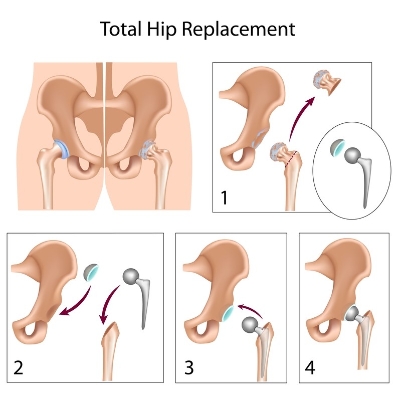
A total hip replacement is performed by removing the top of the femur and replacing it with a metal stem and a ball sized to suit the individual patient's anatomy. The hip socket (acetabulum) is reamed out and fitted with a new cup and a plastic liner. Component choice is guided by long-term data, favouring stems and cups with a proven track record in the New Zealand, Australian, Swedish, and other international joint registries, tailored to the individual, using both cemented and uncemented components depending on what best suits each patient.
Post-Operative Care
Recovery follows a similar structured path to knee replacement, covering pain relief, blood clot prevention, and a step-by-step return to normal activity.
Antibiotics are routinely given around the time of surgery. A total hip replacement also carries a risk of blood clots, and the right balance of preventive medication is chosen carefully, guided by the American Academy of Orthopaedic Surgeons' guideline on preventing thromboembolic disease in elective hip and knee arthroplasty. Warning signs of a clot, pain or swelling below the knee, or of a pulmonary embolus, chest pain or shortness of breath, should prompt immediate medical attention.
Pain is initially managed through an IV line, moving to oral medication before discharge, alongside stool softeners and anti-nausea medication as needed. Staying mobile both in and out of bed in the first few days is important for recovery, with guided in-bed exercises to help.
The average stay is 2–5 days, some people take a little longer, which is entirely normal. At home, small changes make a real difference, commonly used items within easy reach, clear walkways for a walker if needed, rugs and trip hazards removed, a higher chair with armrests, and a chair in the shower.
Driving generally isn't recommended until around 6 weeks, and sexual activity should also be avoided during this time. Sleeping is best done on your back with legs slightly apart, a pillow between them can help. Walking is encouraged as much as comfortable, along with swimming once the wound is dry (around 4 weeks) and cycling on a stationary bike. Follow-up appointments are typically at 2 weeks, 6 weeks, 6 months, and periodically thereafter. Most people are close to their best by 6 months, though some variation between individuals is entirely normal; pain persisting beyond a year is worth fully investigating.
Do: stay active; kneel on the operated leg if needed; keep your leg in front of you when getting up.
Don't: cross your legs at the knees; bend your hip past 90 degrees; sit on low chairs; pick things up off the floor; stop doing your exercises.
A replaced hip is a mechanical device, it's yours to use as you like, but it can wear out, and the more it's used, the sooner that happens. Modern plastic liners are far more durable than older versions, but no sport is generally recommended for the first 6 months, with a gradual, individual approach after that based on what you're hoping to get back to.
Complications
Complications are an inevitable possibility with an operation of this scale. Thankfully they are rare, and as a surgeon I audit my own outcomes to keep them at or below international levels.
- Infection: 0.4–1%
- Blood clot: 0.53%
- Fracture: 0.1–1%
- Dislocation: 1%
- Leg length discrepancy
- Loosening of implant: 1% per year
- Bleeding
- Ongoing pain
- Nerve injury: 0–3%
- Vascular injury: 0.2–0.3%
- Heterotopic ossification
Treatment
Knee Replacement
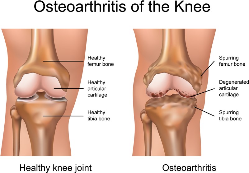
Knee osteoarthritis, the gradual wearing away of the cartilage covering the bones of the knee, is a common condition. As bone begins to rub on bone, it becomes painful, typically felt on the inside of the knee but sometimes on both sides or at the front. Pain is often worse in the mornings, after activity, and on stairs, and the knee can become swollen, stiff, and difficult to fully straighten or bend. Non-operative management, anti-inflammatories, weight management, and activity modification, can help in the earlier stages, though it does not change the underlying course of the arthritis.
The Procedure
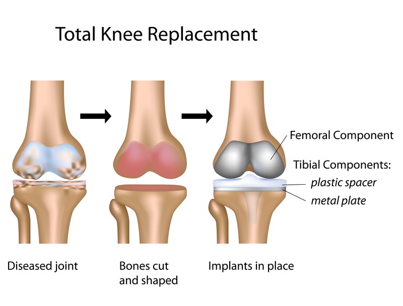
Knee replacement is performed through an incision at the front of the knee. The worn surfaces at the end of the thigh bone and top of the shin bone are replaced with metal components, with a plastic liner between them. Whether the kneecap is also resurfaced depends on individual factors. Most patients stay in hospital for 2–5 days, with physiotherapy starting on the day of surgery. The knee typically continues to improve for up to a year after surgery.
Post-Operative Care
Recovery follows a structured path from hospital through to a full return to activity, covering pain relief, blood clot prevention, and a step-by-step path home.
Antibiotics are routinely given around the time of surgery to reduce infection risk. A total knee replacement also carries a risk of blood clots, and the right balance of preventive medication is chosen carefully, stronger prophylaxis lowers clot risk but increases the chance of bleeding complications. This is guided by the American Academy of Orthopaedic Surgeons' guideline on preventing thromboembolic disease in elective hip and knee arthroplasty. Warning signs of a clot, pain or swelling below the knee, or of a pulmonary embolus, chest pain or shortness of breath, should prompt immediate medical attention.
Pain is initially managed through an IV line, moving to oral medication before discharge, alongside stool softeners and anti-nausea medication as needed. The first few days can feel uncomfortable, and it's important to stay mobile both in and out of bed to support recovery, with guided in-bed exercises to help.
The average stay is 2–5 days, some people take a little longer, which is entirely normal. Getting up and moving independently as early as possible lowers the chance of complications. At home, small changes make a real difference, commonly used items within easy reach, clear walkways for a walker if needed, rugs and trip hazards removed, a higher chair with armrests, and a chair in the shower.
Walking is encouraged as much as comfortable, along with swimming once the wound is dry (around 4 weeks) and cycling on a stationary bike. Follow-up appointments are typically at 2 weeks, 6 weeks, 6 months, 1 year, 2 years, and periodically thereafter. Pain generally settles by 6 weeks, with the knee feeling freer by 6 months, though it does not reach its maximum improvement until around 1 year. Some variation between individuals is entirely normal; pain persisting beyond 2 years is worth fully investigating.
A replaced knee is a mechanical device, it's yours to use as you like, but it can wear out, and the more it's used, the sooner that happens. Regular high-impact activity such as running or tennis is generally not recommended, though the occasional game is fine. Cycling and swimming are good ways to stay fit without the same wear.
Complications
Complications are an inevitable possibility with an operation of this scale. Thankfully they are rare, and as a surgeon I audit my own outcomes to keep them at or below international levels.
- Infection: 0.4–1%
- Blood clot: 1.36%
- Fracture
- Loosening of implant
- Patellofemoral problems (kneecap)
- Bleeding
- Ongoing pain: 19%*
- Peroneal nerve palsy
- Vascular injury
- Stiffness
- Knee instability
*RB Borne et al. Patient satisfaction after total knee arthroplasty: Who is satisfied and who is not. Clin Orthop Relat Res. 2010 Jan;468(1):57-63.
Knee Arthroscopy
For soft tissue problems within the knee, from a locking sensation caused by a meniscal tear to general aching in early degeneration, knee arthroscopy is a relatively small, quick procedure that allows both diagnosis and treatment at the same time. It's performed as day surgery through a couple of small incisions, using a fibre-optic scope to directly visualise and address the problem, with most patients going home the same day on crutches.